CIIDI Distinguished Lecture SERIES
The Center of Excellence in Inflammation, Infectious Diseases, and Immunity emphasizes its interprofessional relationships by hosting annual Speakers Series events. The CIIDI invites all interested parties, including faculty, students, and staff, to informative guest lectures to bring together a diverse body of researchers for both informational purposes and collaborative opportunities.
Upcoming Events
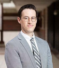
Save the Date: Invited Speaker, Dr. Kevin Byram/CIIDI Member Meeting
|
|
Celebrating a decade of research excellence.
|
Celebrating a decade of research excellence.
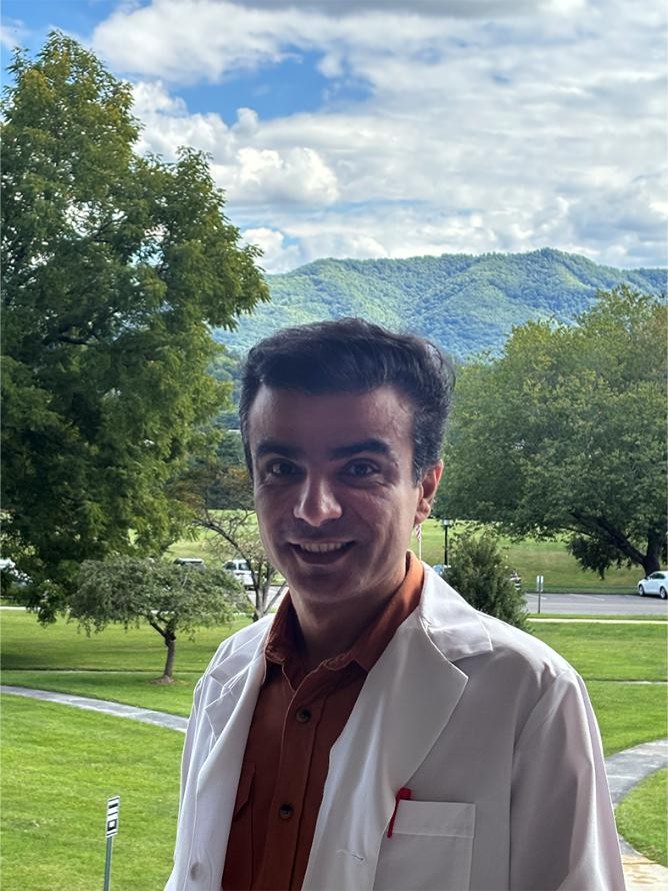
New Faculty Seminar Mohammad Hajipour, PhD"Nanotechnology-Based Approaches for Disease Diagnosis and Therapy: Challenges and Opportunities" October 1, 2025 Bishop Hall, VA 60; Ground Floor Lecture Hall Lunch Served at 11:45 AM; Seminar 12 PM-1 PM |
Past Speakers
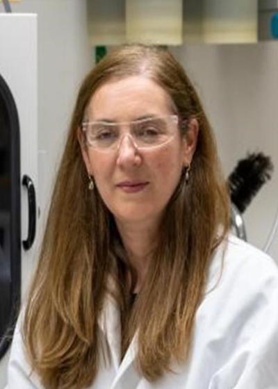
Dr. Sandra Davern, PhD; Section Head, Radioisotope Research and Development Section Oakridge National Laboratory
-
Radiopharmaceuticals: Opportunities for Science and Economic Development
-
Dr. Davern elaborated on the mission of the Oak Ridge National Laboratory, which is to deliver scientific discoveries and technical breakthroughs that will accelerate the development and deployment of solutions in clean energy and global security while creating economic opportunities for the nation.
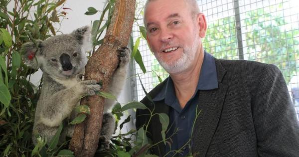
Dr. Peter Timms, PhD; University of Sunshine Coast, Queensland, Australia
-
Development of a Chlamydia Vaccine for Koalas
-
Chlamydia affects up to 50 percent in many of the remaining koala populations, causing blindness, infertility and sometimes death in koalas, consequently hampering population stability and survival.
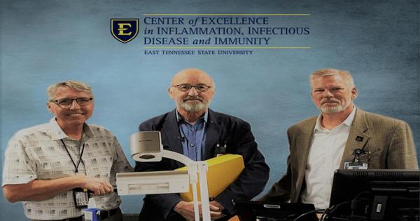
Dr. Robert Davis, MD, MPH
-
The UTHSC 100K Genomes Project
-
Progress in genomic medicine threatens to leave behind medically underserved communities, including the predominantly African American population in west Tennessee as well as the Appalachian population in east Tennessee. This talk will focus on the UTHSC 100K Genomes Project, which is a statewide initiative to study how genomic variants affect health and disease. We currently have over 32,000 subjects enrolled along with exome sequence and SNP data, linked to electronic medical record data, on over 13,000. I will review studies that are currently underway in asthma, epilepsy, and cardiomyopathy and would welcome a discussion of how ETSU researchers might participate and lead studies in their own fields of interest.
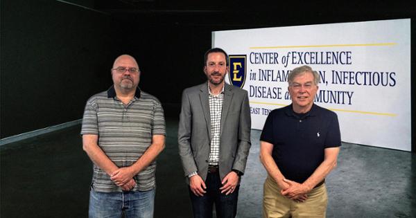
Dr. Brian Peters, PhD
-
Starch or no starch? Ironing out the wrinkles of glycogen metabolism and content in the fungal pathogen Candida albicans
-
Glycogen is a highly branched polymer of glucose and is used across the tree of life as an efficient and compact form of energy storage. While glycogen metabolism pathways have been studied in model yeast, they have not been extensively explored in pathogenic fungi. Using a combination of microbiologic, molecular genetic, and biochemical approaches we reveal orthologous function of glycogen metabolism genes in the fungal pathogen Candida albicans and their deletion leads to fitness and virulence consequences in vivo and unexpected altered host responses via modification of the fungal cell wall.
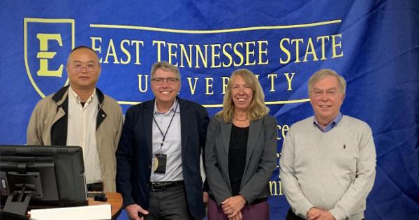
Dr. Jennifer Loftis, PhD
-
Comparison of HCV and SARS-CoV-2 infections on brain function and immune responses
-
Glycogen is a highly branched polymer of glucose and is used across the tree of life as an efficient and compact form of energy storage. While glycogen metabolism pathways have been studied in model yeast, they have not been extensively explored in pathogenic fungi. Using a combination of microbiologic, molecular genetic, and biochemical approaches we reveal orthologous function of glycogen metabolism genes in the fungal pathogen Candida albicans and their deletion leads to fitness and virulence consequences in vivo and unexpected altered host responses via modification of the fungal cell wall.
 Stout Drive Road Closure